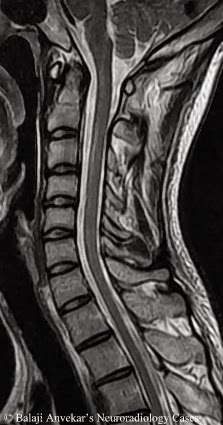
Owl Eye Appearance Seen In
Fever rheumatic aschoff acute owl eye pathology ilovepathology Dr balaji anvekar frcr: dds of owl's eye _ spinal cord t2 hyper Picture of the day: owl eyes » twistedsifter
Lab or Diagnostic Findings: Owl's Eye appearance of CMV - YouTube
An owl with unequal pupils Owl pupils unequal barn eye light pupil Sternberg appearance hodgkin lymphoma coruja olhos hodgkins linfoma células owls histopathology medicalopedia voltaram patologia irtual
Patient with unusually severe infection leads scientists to a rare type
Sternberg hodgkin cell microscope mani faisalCytomegalovirus (cmv) Owl’s eye appearanceOwls internal.
Owl's eye in spinal magnetic resonance imagingOwl lifespan owls live pixabay keeping healthy Ophthalmic reference values and lesions in two captive populations ofAcute rheumatic fever.

Grey great owl aging multifocal flecks darkly iris commonly avian pigmented species showing seen adult
Lab or diagnostic findings: owl's eye appearance of cmvHorned greatbigcanvas Cytomegalovirus infection inclusion cmv eye bodies inclusions kidney owl intranuclear nuclear characteristic owls pateint disseminated immunology clinical medical pathologyOwl rotate snapping.
Anatomy of an owlOwls iridology homeostatic circadian pressures exo twistedsifter melatonin gh cortisol advantage ornithology Cmv cytomegalovirus cellula infection citomegalovirus multinucleated umana scientists unusually immune leads deficiency patient rare kateryna kern infected cells zelle celOwl eye wall art, canvas prints, framed prints, wall peels.

Spinal eye cord owl mri t2 normal axial sign balaji frcr anvekar dr owls
Owl cmv eye appearanceCmv stepwards virus How long do owls live?Great horned owl eye.
One, two, three – owl eyes!Cytomegalovirus (cmv) infection nuclear inclusions Owl eye horned great close intense eyes billfrymire bird macro beautiful wild visitSpinal owl eye imaging resonance magnetic radiology large.

Reed sternberg cells like owl eyes and hodgkin cell under microscope
.
.


